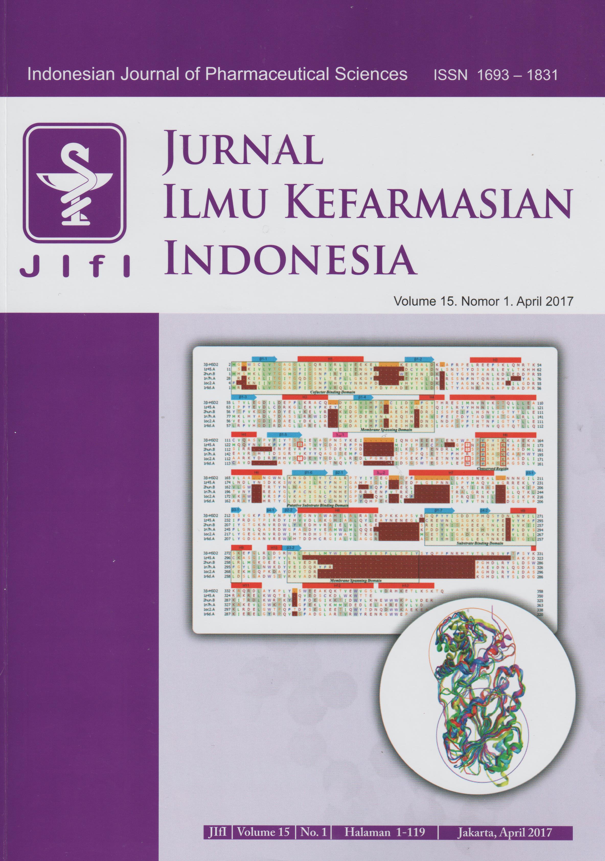
Molecular Modeling of Human 3β-Hydroxysteroid Dehydrogenase Type 2: Combined Homology Modeling, Docking and QSAR Approach
Abstract
A homology model of human 3β-HSD type 2 has been developed from homology modeling techniques using Phyre2 server and refi ned by ModRefi ner. The PROCHECK, QMEAN and ProSA-web online tools were carried out to evaluate the stereochemical quality of the model. The Ramachandran plot resulted from PROCHECK showed that 84.5% residues are in the most favored region, 13.7% are in the additional allowed region, 1.5% are in the generously allowed region and 0.3% are in the disallowed region. The QMEAN (Z-score) are 0.509 (-3.006) and Z-score of ProSA-web is -7.10. The negative values of protein fold energies also found in almost all sequences. Furthermore, molecular docking was also applied to validate the model using MOE. The hydrogen bonding interactions with Tyr154, Ser124, and Ser218 are found in all docked substrates as well as known inhibitors (trilostane and epostane). A dataset of azasteroid inhibitors were also docked into the substrate active site of human 3β-HSD2. These docked structures were utilized to construct corresponding docking-based QSAR equation by employing genetic algorithm (GA) statistical analysis. The contructed best QSAR equation has a robust predictive power according to its statistical parameters, hence may be applied to supersede the default scoring function provided by docking software. These results indicate that the human 3β-HSD2 model was successfully evaluated as a good model.
References
2. Lachance Y, Luu-The V, Verreault H, Dumont M,Rhéaume E, Leblanc G, Labrie F. Structure of the
human type II 3β-hydroxysteroid dehydrogenase/ Δ5-Δ4 isomerase (3β-HSD) gene: adrenal and gonadal specifi city. DNA Cell Biol. 1991.10:701–11. doi:10.1089/dna.1991.10.701.
3. Pennin g TM. Mo lecular endocrinology of hydroxysteroid dehydrogenases. Endocr Rev. 1997.18:281–305. doi:10.1210/edrv.18.3.0302.
4. Simard J, Ricketts ML, Gingras S, Soucy P, Feltus FA, Melner MH. Molecular biology of the 3β-hydroxysteroid dehydrogenase/Δ5-Δ4 isomerase gene family. Endocr Rev. 2005.26:525–82. doi:10.1210/er.2002-0050.
5. Rabbani B, Mahdieh N, Haghi Ashtiani MT, Setoodeh A, Rabbani A. In silico structural, functional and pathogenicity evaluation of a novel mutation: An overview of HSD3B2 gene mutations. Gene. 2012.503:215–21. doi:10.1016/j.gene.2012.04.080.
6. Thomas JL, Duax WL, Addlagatta A, Kacsoh B, Brandt SE, Norris WB. Structure/function aspects of human 3β-hydroxysteroid dehydrogenase. Mol Cell Endocrinol. 2004.215:73–82. doi:10.1016/j. mce.2003.11.018.
7. Goswami AM. Structural modeling and in silico analysis of non-synonymous single nucleotide polymorphisms of human 3β-hydroxysteroid dehydrogenase type 2. Meta Gene. 2015.5:162-72.
8. Apweiler R, Bairoch A, Wu CH, Barker WC,Boeckmann B, Ferro S, et al. UniProt: The universal protein kno wledgebase. Nucleic Acids Res. 2004.32:D115–9. doi:10.1093/nar/gkh131.
9. Berman, H M., Westbrook J, Feng Z, Gilliland G, Bhat, T N, Weissig H, et al. The protein data bank. Nucleic Acids Res. 2000.28:235–42. doi:10.1093/nar/28.1.235.
10. Gaulton A, Bellis LJ, Bento AP, Chambers J, Davies M, Hersey A, Light Y, McGlinchey S, Michalovich D, Al-Lazikani B, Overington JP. ChEMBL: a large-scale bioactivity database for drug discovery. Nucleic Acids Res. 2012.40:D1100–7. doi:10.1093/nar/gkr777.
11. Frye SV, Haff ner CD, Maloney PR, Mook RA Jr, Dorsey GF Jr, Hiner RN, et al. 6-Azasteroids: structureactivity relationships for inhibition of type 1 and 2 human 5 alpha-reductase and human adrenal 3 betahydroxy-delta 5-steroid dehydrogenase/3-keto-delta 5-steroid isomerase. J Med Chem. 1994.37(15):2352- 60.
12. Kelley LA, Mezulis S, Yates CM, Wass MN, Sternberg MJE. The phyre2 web portal for protein modeling, prediction and analysis. Nat Protoc. 2015.10:845–58. doi:10.1038/nprot.2015.053.
13. Molecular Operating Environment (MOE). Canada: Chemical Computing Group Inc; 2015.10.
14. Jones P, Binns D, Chang H-Y, Fraser M, Li W, McAnulla C, et al. InterProScan 5: genome-scale protein function classifi cation. Bioinformatics btu031. 2014. doi:10.1093/bioinformatics/btu031.
15. de Castro E, Sigrist CJA, Gattiker A, Bulliard V, Langendijk-Genevaux PS, Gasteiger E, et al. ScanProsite: detection of PROSITE signature matches and ProRule-associated functional and structural residues in proteins. Nucleic Acids Res. 2006.34:W362–5. doi:10.1093/nar/gkl124.
16. Corpet F, Servant F, Gouzy J, Kahn D. ProDom and ProDom-CG: tools for protein domain analysis and whole genome comparisons. Nucleic Acids Res. 2000.28:267–9. doi:10.1093/nar/28.1.267.
17. Sillitoe I, Lewis TE, Cuff A, Das S, Ashford P, Dawson NL, et al. CATH: comprehensive structural and functional annotations for genome sequences. Nucleic Acids Res. 2015.43:D376–81. doi:10.1093/ nar/gku947.
18. Finn RD, Bateman A, Clements J, Coggill P, Eberhardt RY, Eddy SR, et al. Pfam: the protein families database. Nucleic Acids Res. 2014.42:D222-30.doi:10.1093/ nar/gkt1223.
19. Attwood TK. The PRINTS database: A resource for identifi cation of protein families. Brief Bioinform. 2002.3:252–63.doi:10.1093/bib/3.3.252.
20. Gough J, Karplus K, Hughey R, Chothia C. Assignment of homology to genome sequences using a library of hidden Markov models that represent all proteins of known structure. J Mol Biol. 2001.313:903–19. doi:10.1006/jmbi.2001.5080.
21. Söding J. Protein homology detection by HMM-HMM comparison. Bioinformatics. 2005.21(9):2144.
22. Jeff erys BR, Kelley LA, Sternberg MJE. Protein folding requires crowd control in a simulated cell. J Mol Biol. 2010.397:1329–38.doi:10.1016/j.jmb.2010.01.074.
23. Xu D, Zhang Y. Improving the physical realism and structural accuracy of protein models by a twostep atomic-level energy minimization. Biophys J. 2011.101:2525-34.
24. Labute P. Protonate3D: Assignment of ionization states and hydrogen coordinates to macromolecular structures. Proteins. 2009.75:187–205.doi:10.1002/ prot.22234.
25. Labute P. LowMo deMD: Implicit low mo de velocity fi ltering applied to conformational search of macrocycles and protein loops. J Chem Inf Model. 2010.50:792–800.
26. Laskowski RA, MacArthur MW, Moss D, Thornton JM. PROCHECK: a program to check the stereochemical quality of protein structures. J Appl Cryst. 1993.26:283- 91.
27. Wiederstein M & Sippl MJ. ProSA-web: interactive web service for the recognition of errors in threedimensional structures of proteins. Nucleic Acids Research. 2007.35:W407-10.
28. Jones DT. Protein secondary structure prediction based on position-specifi c scoring matrices. J Mol Biol. 1999.292(2):195-202.
29. Ward JJ, Sodhi JS, McGuffi n LJ, Buxton BF, Jones DT. Prediction and functional analysis of native disorder in proteins from the three kingdoms of life. J Mol Biol. 2004.337(3):635-45.
30. Jones DT. Improving the accuracy of transmembrane protein topology prediction using evolutionary information. Bioinformatics. 2007.23(5):538-44.
31. Kallberg Y, Oppermann U, Persson B. Classifi cation of the short-chain dehydrogenase/reductase superfamily using hidden Markov models. FEBS J. 2010.277:2375- 86.doi:10.1111/j.1742-4658.2010.07656.x.
32. Benkert P, Biasini M, Schwede T. Toward the estimation of the absolute quality of individual protein structure models. Bioinformatics. 2011.27(3):343-50.
33. Bashton M, Chothia C. The geometry of domain combination in proteins. J Mol Biol. 2002.315:927-39. PUBMED:11812158EPMC:11812158.
Licencing
All articles in Jurnal Ilmu Kefarmasian Indonesia are an open-access article, distributed under the terms of the Creative Commons Attribution-NonCommercial-ShareAlike 4.0 International License which permits unrestricted non-commercial used, distribution and reproduction in any medium.
This licence applies to Author(s) and Public Reader means that the users mays :
- SHARE:
copy and redistribute the article in any medium or format - ADAPT:
remix, transform, and build upon the article (eg.: to produce a new research work and, possibly, a new publication) - ALIKE:
If you remix, transform, or build upon the article, you must distribute your contributions under the same license as the original. - NO ADDITIONAL RESTRICTIONS:
You may not apply legal terms or technological measures that legally restrict others from doing anything the license permits.
It does however mean that when you use it you must:
- ATTRIBUTION: You must give appropriate credit to both the Author(s) and the journal, provide a link to the license, and indicate if changes were made. You may do so in any reasonable manner, but not in any way that suggests the licensor endorses you or your use.
You may not:
- NONCOMMERCIAL: You may not use the article for commercial purposes.
This work is licensed under a Creative Commons Attribution-NonCommercial-ShareAlike 4.0 International License.


 Tools
Tools


